Myocardial Infusion Imaging
A widely used diagnostic procedure for heart health is the imaging of the blood infusion to the heart using a low dose of a radioactive tracer in the blood. A large radiation detector rotates about the chest of the patient, detecting gamma radiation from the tracer element which has been injected into the patient's vein. The image shows the perfusion of the blood into the heart muscle.
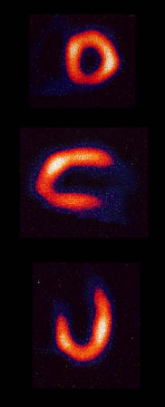 |
The detection of the gamma rays produces images of the blood flow in the heart muscle from various angles to provide an assessment of the infusion of blood. 探测伽马射线可以从不同角度显示心肌中的血流,以评估血液的灌注情况。
Early tests of this type used an isotope of thallium with a gamma ray of energy about 70 keV. This kind of image came to be known as a "thallium scan", but now an isotope of technetium is the isotope of choice for the scans, having a shorter halflife and producing a gamma of energy about 140 keV. 早期这类试验使用一种钍的同位素,其伽马射线的能量约为70 keV。这种图像后来被称为“钍扫描”,但现在更常用的是锝的同位素,因为它半衰期更短,产生的伽马射线能量约为140 keV。
|
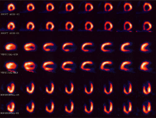
The large detector uses an ionization detection in a manner similar to a Geiger counter and collects a series of exposures as it sequences around the body trunk. A collection of images is illustrated below, taken before and after exercise on a treadmill for comparison of the infusion of the blood under those two conditions.
Nuclear Medicine
| HyperPhysics***** Nuclear
HyperPhysics ***** 核物理 | R Nave |