Bilingual edition: English is preserved and Chinese follows each unit. Terminology uses the confirmed v20260916 glossary; automated semantic review remains traceable.
中英双语版:英文原文完整保留,中文紧随对应单元;术语采用 v20260916 确认表,自动语义审校结果可追溯。

The Retina

视网膜

The retina is a light-sensitive layer at the back of the eye that covers about 65 percent of its interior surface. Photosensitive cells called rods and cones in the retina convert incident light energy into signals that are carried to the brain by the optic nerve. In the middle of the retina is a small dimple called the fovea or fovea centralis. It is the center of the eye's sharpest vision and the location of most color perception.
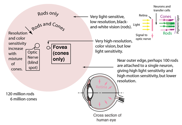
视网膜是位于眼球后方的一个感光层,覆盖了眼球内部表面的约65%。视网膜中含有一种称为杆细胞和锥细胞的感光细胞,它们将入射的光能转化为信号,这些信号通过视神经传递到大脑。视网膜中央有一个小凹陷,称为视斑或中央视斑。它是眼睛最清晰的视觉区域,也是大部分颜色感知的位置。

"A thin layer (about 0.5 to 0.1mm thick) of light receptor cells covers the inner surface of the choroid. The focused beam of light is absorbed via electrochemical reaction in this pinkish multilayered structure. The human eye contains two kinds of photoreceptor cells; rods and cones. Roughly 125 million of them are intermingled nonuniformly over the retina."(Hecht) The ensemble of rods(each about 0.002 mm in diameter) forms an exceedingly sensitive detector, performing in light too dim for the cones to respond to. It is unable to distinquish color, and the images it relays are not well defined.

一层薄薄的光感受细胞(厚度约0.5到0.1毫米)覆盖在脉络膜的内表面。聚焦的光束通过电化学反应在这一粉红色的多层结构中被吸收。人眼含有两种光感受细胞:杆细胞和锥细胞。它们大约有1250万个,非均匀地分布在视网膜上。这些杆细胞(每个直径约0.002毫米)共同形成一个极其敏感的探测器,能够感知锥细胞无法响应的微弱光线。它无法区分颜色,所传递的图像也不清晰。

"In contrast, the ensemble of 6 or 7 million cones (each about 0.006 mm in diameter) can be imagined as a separate, but overlapping, low-speed color film. It performs in bright light, giving detailed colored views, but is fairly insensitive at low light levels."(Hecht)"

相比之下,600万到700万个锥细胞(每个大约0.006毫米直径)可以被想象成一张独立但重叠的低速彩色胶片。它在明亮光线下工作,能提供详细的彩色视图,但对低光水平相对不敏感。

The rods and cones appear to be pointed away from the light source they detect. The light strikes the sensitive pigment layer at the back of the eye and transfers the information to the nearby rods and cones. The rods and cones absorb the scattered light from the nearby pigment layer, which makes vision clearer and keeps the sensitive detectors in a more protected location. The rods and cones are also kept closer to their metabolic energy supply. From the rods and cones, the signals pass through horizontal cells, bipolar cells, amacrine cells and retinal ganglion cells on their way to the retinal highway leading to the optic nerve. (Mammoth)

杆细胞和锥细胞似乎远离它们检测的光源。光击中视网膜后方的敏感色素层,并将信息传递给附近的杆细胞和锥细胞。这些杆细胞和锥细胞吸收来自附近色素层的散射光,使视觉更清晰,并将敏感的探测器置于更安全的位置。杆细胞和锥细胞也更接近它们的代谢能量供应。从杆细胞和锥细胞开始,信号通过水平细胞、双极细胞、视网膜内侧细胞和视网膜神经节细胞,最终通过视网膜高速公路传递到视神经。(Mammoth)
Index

Vision concepts

Image formation concepts

Reference
Hecht, 2nd Ed.
Sec. 5.7

Mammoth Memory site
索引视觉概念 图像形成概念 参考Hecht,第二版第5.7节 Mammoth Memory网站
 
HyperPhysics***** Light and Vision
HyperPhysics ***** 光与视觉
R Nave
Go Back
返回


The Fovea Centralis

中央小孔

Though the eye receives data from a field of about 200 degrees, the acuity over most of that range is poor. To form high resolution images, the light must fall on the fovea, and that limits the acute vision angle to about 15 degrees. In low light, this fovea constitutes a second blind spot since it is exclusively cones which have low light sensitivity. At night, to get most acute vision one must shift the vision slightly to one side, say 4 to 12 degrees so that the light falls on some rods.

尽管眼睛接收来自约200度视野的数据,但大部分范围内视力较差。要形成高分辨率图像,光线必须落在黄斑区,这将视敏度角度限制在约15度。在低光条件下,黄斑区构成第二个盲区,因为只有视锥细胞具有低光灵敏度。夜间,为了获得最清晰的视觉,必须将视线稍微偏移,例如4到12度,使光线落在一些视杆细胞上。

A "dimple on the retina" provides our highest resolution vision.

视网膜上的一个小凹陷提供了我们最清晰的视觉分辨率。

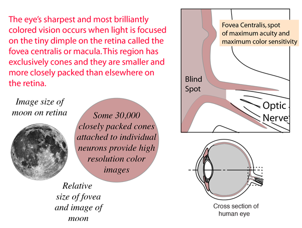
"Just about at the center of the retina is a small depression from 2.5 to 3 mm in diameter known as the yellow spot, or macula. There is a tiny rod-free region about 0.3mm in diameter at its center, the fovea centralis. (In comparison the image of the full Moon on the retina is about 0.2 mm in diameter.) Here the cones are thinner (with diameters of 0.0030mm to 0.0015mm) and more densely packed than anywhere else in the retina. Since the fovea provides the sharpest and most detailed information, the eyeball is continuously moving, so that light from the object of primary interest falls on this region. ...the rods are multiply connected to nerve fibers, and a single such fiber can be activated by any one of about a hundred rods. By contrast, cones in the fovea are individually connected to nerve fibers. The actual perception of a scene is constructed by the eye-brain system in a continuous analysis of the time-varying retinal image."(Hecht)

在视网膜中心附近有一个直径从2.5到3毫米的小凹陷,称为黄斑或视锥区。其中心有一个直径约0.3毫米的微小无杆区,称为中央窝。(相比之下,满月在视网膜上的影像直径约为0.2毫米。)在这里,视锥细胞更细(直径为0.0030毫米到0.0015毫米)且密度更高,比视网膜其他任何地方都高。由于中央窝提供最清晰和最详细的信息,眼睛持续在移动,使得物体的主要影像落在这一区域。……视杆细胞与神经纤维多重连接,一根这样的纤维可以被约一百个视杆细胞激活。相比之下,中央窝的视锥细胞则单独连接到神经纤维。实际的场景感知是由眼-脑系统持续分析时间变化的视网膜影像而构建的。
Index

Vision concepts

References
Hecht, 2nd Ed.
Sec. 5.7
Williamson & Cummins
索引 视觉概念 参考文献 海特第二版 第5.7节 Williamson & Cummins
 
HyperPhysics***** Light and Vision
HyperPhysics ***** 光与视觉
R Nave
Go Back
返回


Optic Nerve

视神经

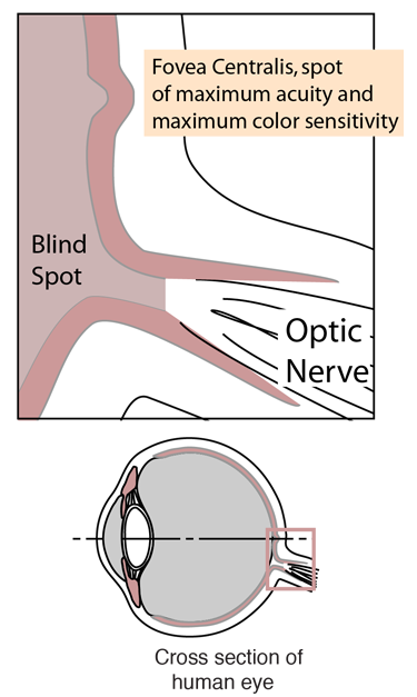
The optic nerve is the cable of nerve fibers with carries the electrical signals from the retina to the brain for processing. The point of departure of that optic nerve through the retina does not have any rods or cones, and thus produces a "blind spot".

视神经是由神经纤维组成的电缆,负责将视网膜上的电信号传递至大脑进行处理。视神经从视网膜穿出的部位没有视杆细胞或视锥细胞,因此形成一个‘盲点’。
Index

Vision concepts

References
Hecht, 2nd Ed.
Sec. 5.7
Williamson & Cummins
索引 视觉概念 参考文献 海特第二版 第5.7节 Williamson & Cummins
 
HyperPhysics***** Light and Vision
HyperPhysics ***** 光与视觉
R Nave
Go Back
返回