Bilingual edition: English is preserved and Chinese follows each unit. Terminology uses the confirmed v20260916 glossary; automated semantic review remains traceable.
中英双语版:英文原文完整保留,中文紧随对应单元;术语采用 v20260916 确认表,自动语义审校结果可追溯。

Nerve Cell

神经细胞

In the body's nervous system are many nerve cells are of the basic type illustrated above. Some kind of stimulus triggers an electric discharge of the cell which is analogous to the discharge of a capacitor. This produces an electrical pulse on the order of 50-70 millivolts called an action potential. The electrical impulse propagates down the fiber-like extension of the nerve cell (the axon). The speed of transmission depends upon the size of the fiber, but is on the order of tens of meters per second - not the speed of light transmission that occurs with electrical signals on wires. Once the signal reaches the axon terminal bundle, it may be transmitted to a neighboring nerve cell with the action of a chemical neurotransmitter.

人体的神经系统中有很多神经细胞,其基本类型如上所示。某种类型的刺激会引发细胞内的电放电,这种放电类似于电容器的放电。这会产生一个约50-70毫伏的电信号,称为动作电位。该电信号沿着神经细胞的纤维状延伸部分(即轴突)传播。传导速度取决于纤维的大小,但通常为每秒数十米——这与通过电线传输的电信号的速度不同。一旦信号到达轴突末端,它可能通过化学神经递质传递到相邻的神经细胞。

The dendrites serve as the stimulus receptors for the neuron, but they respond to a number of different types of stimuli. The neurons in the optic nerve respond to electrical stimuli sent by the cells of the retina. Other types of receptors respond to chemical neurotransmitters.

树突作为神经元的刺激受体,但它们对多种不同类型的刺激作出反应。视神经中的神经元对由视网膜细胞发出的电信号作出反应。其他类型的受体则对化学神经递质作出反应。

The cell body contains the necessary structures for keeping the neuron functional. That includes the nucleus, mitochondria, and other organelles. Extending from the opposite side of the cell body is the long tubular extension called the axon. Surrounding the axon is the myelin sheath, which plays an important role in the rate of electrical transmission. At the terminal end of the axon is a branched structure with ends called synaptic knobs. From this structure chemical signals can be sent to neighboring neurons.

细胞体包含保持神经元功能所必需的结构。这包括细胞核、线粒体和其他细胞器。从细胞体的另一侧延伸出一个长管状结构,称为轴突。轴突周围有髓鞘,它在电流传导速度方面起着重要作用。轴突末端是一个带有末端的分支结构,称为突触小体。从该结构可以向邻近神经元发送化学信号。
Transmission of nerve impulse along the axon
轴突中神经冲动的传递

Contributing author: Ka Xiong Charand

贡献作者:Ka Xiong Charand
Index

Bioelectricty
索引 生物电
 
HyperPhysics***** Biology
HyperPhysics ***** 生物学
R Nave
Go Back
返回


Transmission of a nerve impulse along an axon

神经冲动沿轴突的传导

A nerve cell is like a receiver, transmitter and transmission line with the task of passing a signal along from its dendrites to the axon terminal bundle.

神经细胞就像一个接收器、发射器和传输线路,其任务是将信号从树突传递到轴突末梢。

The stimulus triggers an action potential in the cell membrane of the nerve cell, and that action potential provides the stimulus for a neighboring segment of the cell membrane. When the propagating action potential reaches the axon, it proceeds down that "transmission line" by successive excitation of segments of the axon membrane.

刺激引发神经细胞膜中的动作电位,该动作电位为相邻膜段提供刺激。当传播的动作电位到达轴突时,它通过依次激发轴突膜段,沿这条‘传输线路’向下传播。

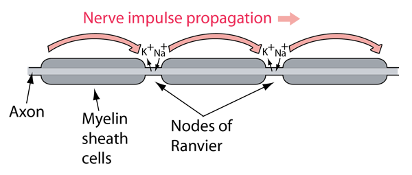
Just the successive stimulation of action potentials would result in slow signal transmission down the axon. The propagation speed is considerably increased by the action of the myelin sheath.

仅仅连续的动作电位刺激会导致信号在轴突内缓慢传递。髓鞘的作用显著提高了信号的传播速度。

The myelin sheath around the axon prevents the gates on that part of the axon from opening and exchanging their ions with the outside environment. There are gaps between the myelin sheath cells known as the Nodes of Ranvier. At those uncovered areas of the axon membrane, the ion exchange necessary for the production of an action potential can take place. The action potential at one node is sufficient to excite a response at the next node, so the nerve signal can propagate faster by these discrete jumps than by the continuous propagation of depolarization/repolarization along the membrane. This enhanced signal transmission is called saltatory conduction (from the Latin saltare, to jump or hop).

轴突周围的髓鞘阻止了该部分轴突上的离子与外界环境进行交换。髓鞘细胞之间的间隙称为瑞吉尔节点。在这些未被覆盖的轴突膜区域,离子交换对于产生动作电位是必要的。一个节点处的动作电位足以激发下一个节点的反应,因此神经信号可以通过这些离散的跳跃方式传播得比通过膜的连续去极化/复极化传播更快。这种增强的信号传递称为跳跃传导(来自拉丁语saltare,意为跳跃或跃步)。

Tuzynski and Dixon offer some quantification of the sizes involved in these nerve cells. The axon is made up of connected segments of length about 2 mm and diameter typically 20 mm. This diameter compares to about 100 mm for the diameter of a human hair. Axon diameters may vary from 0.1 mm to 20 mm and may be up to a meter long. The much-studied squid has a giant axon of about a millimeter in diameter. The myelin sheaths are about 1mm in length. The action potential travels along the axon at speeds from 1 to 100 m/s.

Tuzynski 和 Dixon 对这些神经细胞中涉及的尺寸进行了量化。轴突由长度约为2毫米、直径通常为20微米的连接段组成。这个直径与人类头发直径约100微米相比要小。轴突直径可能从0.1微米到20微米不等,长度可能达到一米。被广泛研究的章鱼具有直径约一毫米的巨轴突。髓鞘的长度约为1毫米。动作电位沿轴突传导的速度范围从1到100米/秒。


Contributing author: Ka Xiong Charand

贡献作者:Ka Xiong Charand
Index

Bioelectricty

Tuzynski & Dixon
Sec 20.2
索引 生物电 Tuzynski & Dixon 第20.2节
 
HyperPhysics***** Biology
HyperPhysics ***** 生物学
R Nave
Go Back
返回


Neural Stimulation of a Muscle Fiber

肌肉纤维的神经刺激

Muscle fibers contract by the action of actin and myosin sliding past each other. The signal to initiate the contraction comes from the brain as a part of the somatic nervous system.

肌纤维通过肌动蛋白和肌球蛋白相互滑动的作用而收缩。收缩的信号来自大脑,作为躯体神经系统的一部分。

The illustration below is a schematic representation of the process from the arrival of a nerve signal to the terminal bundle of the nerve axon to the contration of a muscle fiber. The stimulation of muscle action is associated with the neurotransmitter chemical acetylcholine.
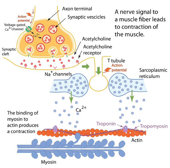
下图是神经信号到达神经轴突末梢到肌肉纤维收缩过程的示意图。肌肉收缩与神经递质乙酰胆碱有关。

When the nerve signal from the somatic nerve system reaches the muscle cell, voltage-dependent calcium gates open to allow calcium to enter the axon terminal. This calcium moves the acetylcholine-containing miceles to fuse with the presynaptic membrane and release their acetylcholine into the synapse, where it is bound by acetylcholine receptors on the postsynaptic surface. The acetylcholine receptors are examples of ligand-gated ion channels: upon binding the acetylcholine molecule, they open up a channel for sodium and potassium ions to enter the cell. In this case acetylcholine is the "ligand" that opens the gate for sodium.

当躯体神经系统的神经信号到达肌肉细胞时,电压依赖的钙门打开,允许钙离子进入轴突末梢。这种钙离子使含有乙酰胆碱的分子与突触前膜融合,释放出乙酰胆碱到突触间隙,其中乙酰胆碱与突触后膜上的乙酰胆碱受体结合。乙酰胆碱受体是配体门控离子通道的例子:当乙酰胆碱分子结合时,它们会打开钠和钾离子的通道。在这种情况下,乙酰胆碱是打开钠离子通道的‘配体’。

When the opening of the Na channels sends a rush of Na into the cell, which, if it is strong enough, causes nearby voltage-gated Na channels to open and produces an action potential. This action potential is not one in a nerve cell, but in the muscle cell.

当钠通道的开启使钠离子涌入细胞时,如果这一过程足够强烈,会引发附近电压门控钠通道的开启,产生动作电位。这种动作电位不是在神经细胞中发生的,而是在肌细胞中发生的。

The muscle fiber structure has lots of tubes called T-tubules or transverse tubules. When the action potential travels down these tubules, it eventually triggers the voltage-sensitive proteins that are linked to the calcium channels in the structure called the sarcoplasmic reticulum (Wiki) that surrounds the nerve fibers. This membrane-enclosed structure has similarities to the endoplasmic reticulum in other cells. In the rest state, the sarcoplasmic reticulum will have a reserved supply of calcium because its walls have many Ca pumps which use ATP energy to store calcium. With the stimulus of the action potential, calcium rushes into the cell and interacts with the actin. Associated with the actin are the troponin complex and the tropomyosin strand which block the binding of myosin. The supplied calcium ions bind to the troponin and pulls the "gaurding" troponin and tropomyosin strand away from the site where myosin can bind.

肌纤维结构中有许多称为T管或横向管的管状结构。当动作电位沿着这些管状结构传播时,最终会触发与结构中的钙通道相连的电压敏感蛋白,该结构称为肌浆网(Wiki),围绕着神经纤维。这种被膜包围的结构在其他细胞中与内质网有相似之处。在静息状态下,肌浆网由于其壁上有许多钙泵,会储存钙离子,这些钙泵利用ATP能量来储存钙。在动作电位的刺激下,钙离子涌入细胞并与肌动蛋白结合。与肌动蛋白结合的还有肌钙蛋白复合物和肌凝蛋白丝,它们会阻止肌球蛋白的结合。提供的钙离子会与肌钙蛋白结合,并将“守卫”的肌钙蛋白和肌凝蛋白丝从肌球蛋白结合的位点移开。

In order to bind to the actin, the myocin must have a supply of energy, which it obtains from ATP . Having absorbed energy from ATP, a unit of the myosin fiber will be in a stressed or high energy state, like a stretched spring. With the action of the calcium to withdraw the troponin and tropomyosin, the myosin structure can bind and use the energy to pull the actin fiber, shortening or contracting the muscle fiber.
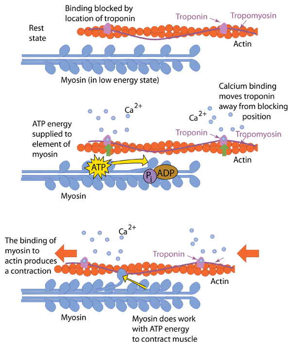
为了与肌动蛋白结合,肌球蛋白必须有能量供应,它通过ATP获取能量。吸收了ATP中的能量后,肌球蛋白纤维中的一个单位将处于紧张或高能状态,就像被拉伸的弹簧一样。在钙的作用下,肌球蛋白结构可以结合并利用能量将肌动蛋白纤维拉拢,使肌肉纤维缩短或收缩。

While the contraction of a muscle can be repeated by following the above steps, there must be a pathway back to a rest state since you don't want your muscles to be in a permanently contracted state. Those mechanisms for return to rest are provided. The initial stimulus by the motor nerve which started the process is under conscious control, so you can decide to relax the muscle. The free acetylcholine in the synaptic gap is removed by another molecule, acetylcholinesterase. This is an essential function in neuromuscular junctions since the continued presence of acetylcholine could keep the muscle in extended contraction. In fact, the role of nerve poisons and the venom of some snakes is to block the action of acetylcholinesterase and force the muscles into a continuing contracted state. In normal operation the acetylcholinesterase works briefly and then ceases. The calcium pumps in the sarcoplasmic reticulum work to reclaim the calcium, and upon removal of the calcium from the receptors on the muscle, the "bodygaurd" troponin and tropomyocin move back to their blocking positions. The myosin and actin fibers return to their relaxed state.

虽然按照上述步骤可以重复肌肉的收缩,但必须有一条途径返回到休息状态,因为你不希望肌肉一直处于永久收缩状态。这些返回休息的机制是存在的。启动这一过程的神经冲动是由运动神经元发出的,受意识控制,因此你可以决定放松肌肉。突触间隙中的游离乙酰胆碱由另一种分子——乙酰胆碱酯酶清除。这一功能在神经肌肉接点中是至关重要的,因为乙酰胆碱的持续存在可能会使肌肉保持持续收缩状态。事实上,某些毒药和蛇的毒液的作用就是阻断乙酰胆碱酯酶的作用,迫使肌肉保持持续收缩状态。在正常运作中,乙酰胆碱酯酶仅短暂起作用后就会停止工作。肌浆网中的钙泵负责回收钙离子,当钙离子从肌肉受体上移除后,‘守卫’分子肌钙蛋白和肌动蛋白会回到阻断位置。肌球蛋白和肌动蛋白纤维则恢复到放松状态。
Index

Bioelectricty

Tuzynski & Dixon
Sec 20.2

Neural Stimulation, lumenlearning

Frontera & Ochala

Acetylcholine receptor, Britannica

BioNinja
索引 生物电 Tuzynski & Dixon 第20.2节 神经刺激,lumenlearning Frontera & Ochala 乙酰胆碱受体,Britannica BioNinja
 
HyperPhysics***** Biology
HyperPhysics ***** 生物学
R Nave
Go Back
返回